


How Modern Imaging Has Improved Heart Disease Detection
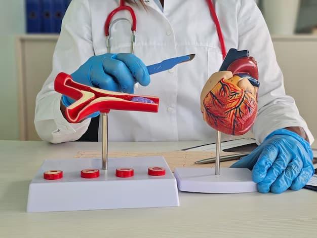
Heart disease remains one of the leading health concerns worldwide, but one of the biggest advancements in cardiology over the past two decades has been the ability to detect heart problems much earlier than before. Earlier, many patients were diagnosed only after suffering a heart attack or developing severe symptoms. Today, modern cardiac imaging allows doctors to identify abnormalities at an earlier stage, helping patients receive timely treatment before serious complications occur.
The ability to look inside the heart and coronary arteries with advanced imaging technologies has transformed the diagnosis and management of cardiovascular diseases. These investigations provide detailed information about heart function, blood flow, valve abnormalities, and coronary artery blockages, allowing cardiologists to create personalized treatment plans based on accurate findings rather than assumptions.
Patients seeking Coronary Angiography Treatment in Jaipur now benefit from advanced diagnostic techniques that make heart disease detection more precise, helping doctors choose the most appropriate treatment while reducing unnecessary delays.
Why Early Detection Makes a Significant Difference
Heart disease often develops silently over many years. Cholesterol deposits gradually build up inside the coronary arteries, reducing blood flow to the heart muscle. During the early stages, patients may not experience any symptoms, or they may notice only mild discomfort that is easy to ignore.
Unfortunately, many people assume that heart disease always causes severe chest pain. In reality, some patients experience only fatigue, breathlessness, reduced exercise capacity, or mild chest heaviness during physical activity. Others may have no symptoms until a major cardiac event occurs.
Early detection helps identify these hidden problems before they become life-threatening. By diagnosing coronary artery disease at the right time, doctors can begin treatment earlier, improve blood flow, control risk factors, and reduce the chances of heart attack or heart failure.
Understanding the Role of Modern Cardiac Imaging
Cardiac imaging refers to a group of advanced diagnostic techniques that allow doctors to evaluate different aspects of heart health.
Instead of relying only on physical examination or symptoms, cardiologists can now assess the heart's structure, pumping function, valve movement, blood circulation, and coronary arteries in remarkable detail.
Each imaging test has a specific purpose. The choice depends on the patient's symptoms, medical history, age, risk factors, and previous investigations. Using the right investigation at the right time helps ensure an accurate diagnosis while avoiding unnecessary procedures.
Electrocardiogram Helps Detect Electrical Changes
One of the first tests performed when a patient presents with chest discomfort or suspected heart disease is an electrocardiogram (ECG).
This simple investigation records the electrical activity of the heart and can identify rhythm disturbances, evidence of previous heart damage, or changes that may suggest reduced blood supply to the heart muscle.
Although an ECG alone cannot detect every heart problem, it provides valuable information that helps guide further evaluation.
Echocardiography Shows How the Heart Is Functioning
Echocardiography uses ultrasound waves to produce moving images of the heart.
This investigation helps doctors assess heart muscle function, pumping efficiency, valve movement, chamber size, and blood flow through different parts of the heart.
It is particularly useful in evaluating patients with breathlessness, heart murmurs, heart failure, and suspected structural heart disease.
Since it does not involve radiation and provides real-time information, echocardiography remains one of the most commonly used cardiac imaging techniques.
Stress Testing Helps Identify Hidden Blood Flow Problems
Some patients experience symptoms only during physical activity because the heart requires more oxygen during exercise.
Stress testing evaluates how the heart responds under increased workload. If reduced blood flow develops during exercise, the test may indicate the presence of coronary artery disease.
Stress testing is often combined with imaging techniques to improve diagnostic accuracy in selected patients.
The results help cardiologists decide whether additional investigations are necessary.
Coronary Angiography Remains the Gold Standard for Detecting Blockages
Among all imaging techniques, coronary angiography continues to be one of the most accurate methods for evaluating the coronary arteries.
During this procedure, a thin catheter is introduced through a blood vessel, usually from the wrist or groin, and guided to the coronary arteries. A special contrast dye is injected while high-resolution imaging captures detailed pictures of blood flow.
Coronary angiography allows doctors to identify the exact location, number, and severity of arterial blockages.
Patients undergoing Coronary Angiography Treatment in Jaipur benefit from precise diagnosis that helps determine whether medications, angioplasty, or other treatment options are most appropriate.
Imaging Helps Differentiate Angina From Other Causes of Chest Pain
Chest pain is one of the most common reasons patients visit a cardiologist, but not every episode of chest discomfort originates from the heart.
Muscle strain, acidity, anxiety, lung disorders, and digestive conditions can all produce symptoms similar to cardiac pain.
Modern imaging helps distinguish coronary artery disease from these other conditions.
Patients requiring evaluation for Angina Treatment in Jaipur often undergo detailed cardiac investigations to determine whether reduced blood flow is responsible for their symptoms. Accurate diagnosis ensures that treatment is directed toward the actual cause rather than simply relieving discomfort.
Similarly, individuals seeking Chest Pain Treatment in Jaipur benefit from comprehensive evaluation that combines clinical assessment with advanced imaging to identify both cardiac and non-cardiac causes.
Better Imaging Leads to Better Treatment Planning
The greatest advantage of modern imaging is not simply detecting disease—it is helping doctors make informed treatment decisions.
Once accurate images are available, cardiologists can determine whether the patient requires lifestyle modification, medications, coronary angioplasty, stent placement, or continued observation.
Rather than following a standard treatment pathway for every patient, imaging allows care to be individualized according to the severity and location of disease.
This personalized approach improves both safety and long-term outcomes.
Technology Has Made Cardiac Procedures Safer
Advances in imaging technology have also improved the safety of interventional cardiology procedures.
High-resolution imaging provides clearer visualization of coronary arteries, allowing greater precision during coronary angiography and angioplasty.
Modern catheter techniques, improved imaging systems, and enhanced procedural planning have contributed to faster recovery, reduced complications, and improved patient comfort.
These technological developments continue to improve the overall quality of cardiovascular care.
Why Regular Heart Evaluation Should Not Be Delayed
Many patients postpone cardiac evaluation because they believe symptoms will disappear on their own or because they fear undergoing investigations.
However, delaying diagnosis may allow coronary artery disease to progress silently.
Individuals with diabetes, hypertension, high cholesterol, obesity, smoking history, family history of heart disease, or persistent chest discomfort should undergo timely cardiac assessment even if symptoms appear mild.
Early imaging can identify disease before major complications develop, allowing treatment to begin when it is most effective.
Advanced Cardiac Imaging by Dr. Rahul Sharma
Accurate diagnosis forms the foundation of successful heart treatment.
Dr. Rahul Sharma is an experienced cardiologist with qualifications including MBBS, DNB (General Medicine), and DNB (Cardiology) and more than 20 years of experience in advanced cardiovascular care.
His expertise includes coronary angiography, coronary angioplasty, preventive cardiology, hypertension management, chest pain evaluation, heart failure management, emergency cardiac care, and comprehensive diagnosis of coronary artery disease.
Patients seeking Coronary Angiography Treatment in Jaipur receive detailed cardiac evaluation using modern diagnostic techniques that help identify coronary blockages accurately and guide appropriate treatment.
Individuals requiring consultation for Angina Treatment in Jaipur or expert Chest Pain Treatment in Jaipur benefit from his evidence-based approach, careful clinical assessment, and personalized treatment plans designed to protect long-term heart health.
Final Thoughts
Modern cardiac imaging has transformed the way heart disease is diagnosed. Instead of waiting for serious symptoms or emergencies, doctors can now identify many cardiovascular problems at much earlier stages, allowing timely intervention and better outcomes.
Whether evaluating chest pain, suspected angina, or coronary artery disease, advanced imaging provides the detailed information needed to make accurate treatment decisions.
By combining early diagnosis, advanced technology, healthy lifestyle choices, and expert cardiac care, patients have a much greater opportunity to prevent complications and enjoy a healthier future with a stronger heart.

 English
 Arabic
 Spanish
 Portuguese
 Deutsch
 Turkish
 Dutch
Dutch
 Italiano
 Russian
 Romaian
 Portuguese (Brazil)
 Greek
Greek